Looking through the PUMA at a tardigrade
Every image on this page is computed, none is painted. Light from a white LED travels as a wave through a real, nanoCT-scanned tardigrade. It then passes lenses whose data come from open patents: the objective, the eyepiece and a model eye. The colour fringes, blur and diffraction you see all come out of that calculation.
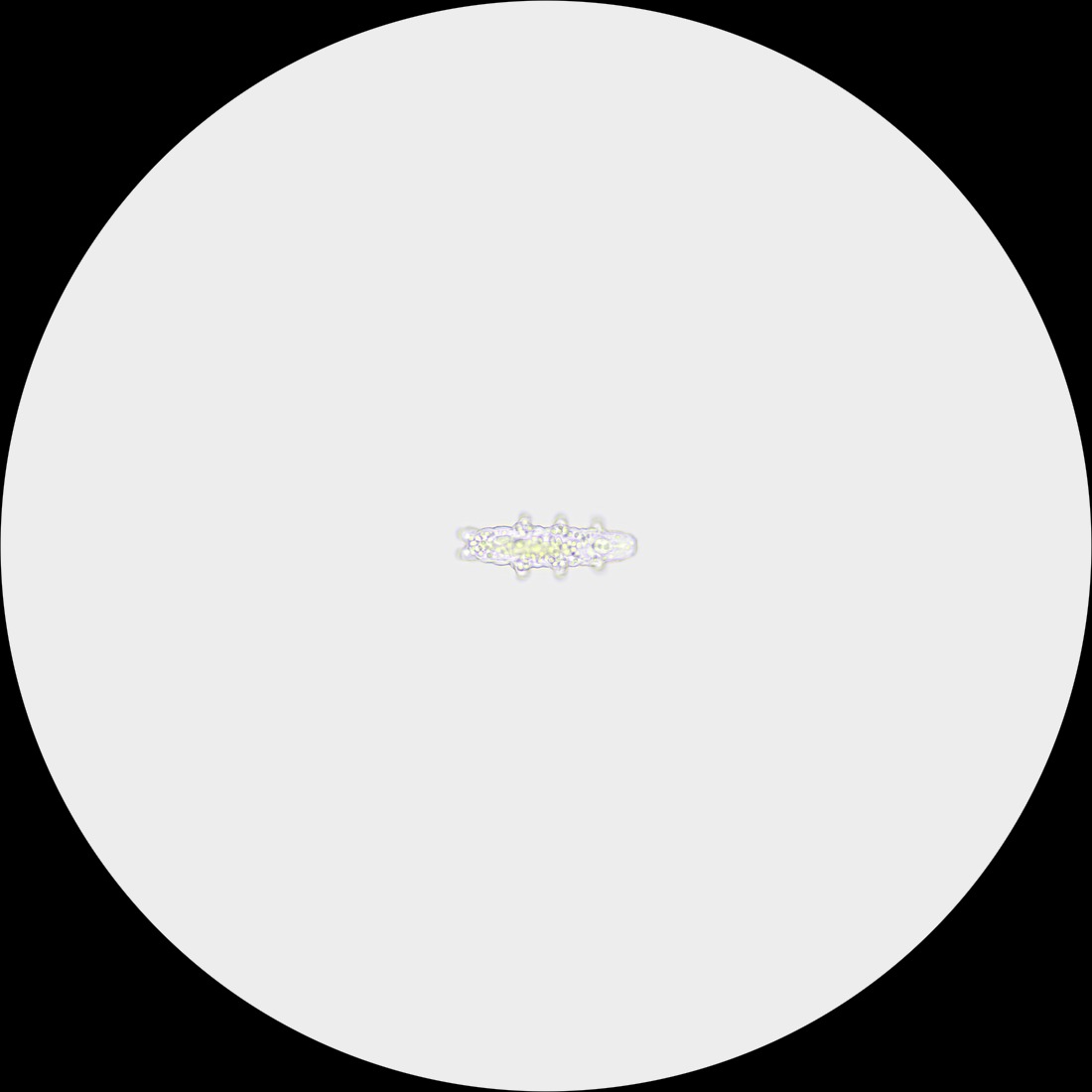
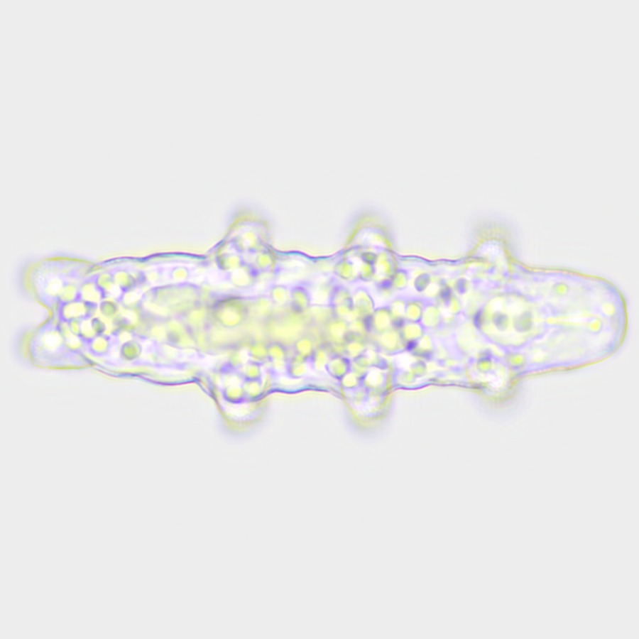
Drag inside the ring: roll the animal · on the ring: turn the slide · wheel, pinch or ↑ ↓: focus · ← →: turn · double-click: back to the start view
The instrument
Drag to rotate, scroll or pinch to zoom. Built from the original PUMA CAD parts (FreeCAD → Blender → glTF), assembled by hand from the PUMA build guide, so part positions are approximate.

The animal walking
The optical path
- White LED, 6500 KPUMA docsBlue InGaN peak at 452 nm plus phosphor. 9 wavelengths from 420 to 680 nm.
- Illumination: mirror or Köhler condenserselectableThe PUMA Foundation scope lights the slide with a plane mirror (modelled as NA ≈ 0.05). From 20x up the PUMA docs call for a condenser (measured up to NA 0.70). The Köhler aperture stop can be set to NA 0.15, 0.30 or 0.40.
- Tardigrade Hypsibius exemplarisnanoCT, CC BY152 µm long, 270 nm voxels, 11 segmented organs. Refractive index per tissue is a literature-based assumption.
- Cover glass 0.17 mm, waterSchott D 263Focusing deeper into the water adds spherical aberration. The calculation includes it.
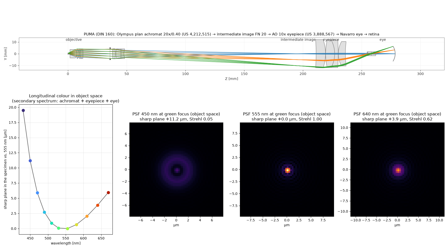
- Plan achromat 20x/0.40Olympus patentUS 4,212,515, 7 elements, glasses S-LAH66, S-FPL51, S-LAL12 and others. A generic objective rather than a specific retail part, as the PUMA intends.
- Tube 160 mm, field stop 20 mmPUMA docsFinite DIN system without a tube lens.
- Eyepiece 10xAO patentUS 3,888,567, f = 24.9 mm. The PUMA only specifies "WF 10x/20".
- EyeNavarro 1985Aspheric model eye with dispersion of the ocular media. The exit pupil is 1.05 mm.
What is computed
- Wave propagation through the 3D animal in 134 slices (multi-slice), with diffraction, refraction and multiple scattering.
- Partially coherent illumination as an incoherent sum over condenser source points (7 to 91, depending on the aperture) × 9 wavelengths.
- Longitudinal colour as a wavelength-dependent sharp plane, found by ray tracing from the specimen to the retina. At 450 nm it lies 11 µm deeper than for green.
- Residual aberrations (spherical aberration, spherochromatism) as a pupil phase from the traced wavefront.
- Colour through the CIE 1931 colour-matching functions, white-balanced like an eye adapted to the lamp.
What is assumed or simplified
- Tissue refractive indices: cytoplasm ≈ 1.355, storage cells ≈ 1.39, cuticle ≈ 1.46. Algae in the gut absorb with a chlorophyll-like spectrum.
- Only the central 207 µm are simulated. The rest of the 1 mm field of view is empty slide in the simulated background colour.
- Aberrations are those on axis. Field curvature towards the edge of the field is not included.
- Scalar wave optics, no polarisation. The detail view is a digital zoom on the same data.
- The scanned animal was critical-point dried and is therefore smaller than a living one (living adults are 250–500 µm).
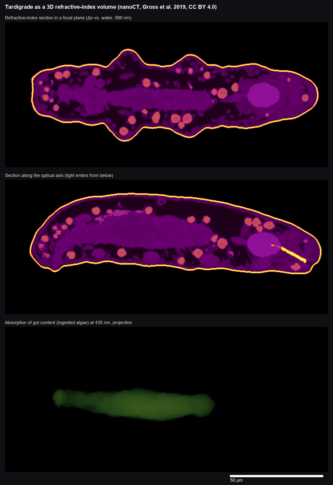
Sources and licences. Tardigrade scan: Gross, V. et al. (2019) "X-ray imaging of a water bear offers a new look at tardigrade internal anatomy", Zoological Letters 5:14, CC BY 4.0. The PUMA microscope was invented and designed by Dr Paul J. Tadrous (@TadPath): github.com/TadPath/PUMA (CAD GPL-3.0, docs GFDL); paper: J. Microsc. 283:259–280 (2021), doi:10.1111/jmi.13043. This page is an independent project built on PUMA and is not endorsed by its author. Objective: US 4,212,515 (Itaya, Olympus 1980). Eyepiece: US 3,888,567 (Shoemaker, American Optical 1975). Eye: Navarro, Santamaría & Bescós, JOSA A 2 (1985). Ray tracing: Optiland (MIT). Glass data: Ohara and Schott catalogues. 3D viewer: model-viewer (Apache-2.0).